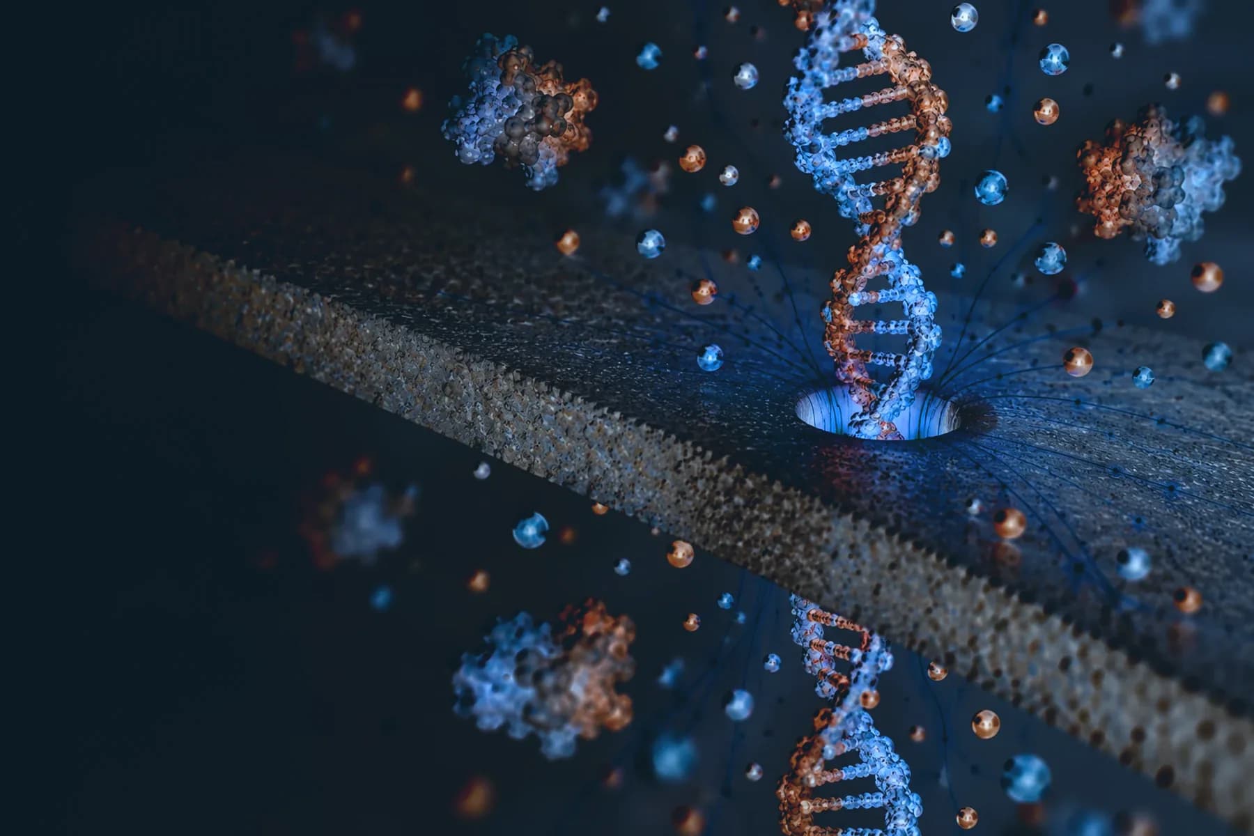
A hole, a voltage and a current
Take a membrane a few nanometres thick, open a single pore in it a few nanometres across, and fill both sides with salt water. Apply a small voltage across the membrane and ions flow through the pore. That flow is an electrical current we can record continuously, and with nothing in the way it is steady.
Now add molecules to one side. When one of them is drawn into the pore, it occupies space that the electrolyte used to fill, so fewer ions can pass and the current drops. When the molecule leaves on the far side, the current recovers. Each passage leaves a short dip in the trace, which we call an event, and a single recording can contain many thousands of them.
Almost everything I do in biosensing starts from this measurement. The rest of this article is about what a dip can tell you, and why so much of my work goes into the membrane rather than the molecule.
What an event tells you
Two numbers describe most events. The first is the depth of the dip. To a first approximation, the drop in current scales with the volume of electrolyte the molecule excludes from the pore, so a larger molecule gives a deeper blockade. Shape matters too: a compact molecule and an elongated one of similar mass need not block the pore in the same way.
The second is the dwell time, how long the dip lasts. It depends on how hard the electric field pulls on the molecule, which is set by the molecule's charge and the applied voltage, and on how much the molecule interacts with the pore walls on its way through. Beyond these two numbers, the current inside the dip has its own structure, and that becomes important once machine learning is involved. I wrote about that in a separate article on identifying proteins.
In my experiments the pore is wider than the molecule being measured. A folded protein or a folded RNA goes through intact, as a compact particle, and its event reflects that folded shape. This is what makes it possible to use a pore to study molecular shape at all, as in our work on a disease-linked tRNA. DNA behaves differently because it is a long, thin polymer: it threads through the pore along its length, and the event lasts as long as the strand takes to pass.
These relationships are approximate. Event signals also depend on the pore, the voltage and the electrolyte. In Langmuir (2024) we compared experiments and simulations of how electrolyte properties change ion transport through solid-state nanopores, because you need that baseline before you can interpret a blockade with any confidence.
Why thin membranes matter
The electrical resistance of a pore is spread along its length. In a thick membrane, a molecule occupies only part of that length at any moment, so the change it causes is diluted. A thinner membrane concentrates the sensing region and makes each blockade easier to resolve.
The catch is that very thin membranes tend to be fragile, and a pore that changes or fails partway through an experiment limits how much data you can collect. In Analytical Chemistry (2023) we reported a scalable route to ultra-thin silicon nitride membranes with an effective thickness of about 3 nm. A single pore in one of these membranes recorded more than 500,000 DNA translocations and 1.8 million protein translocations under the conditions in the paper, and the work was featured on the journal's cover.
Long-lived pores matter because the statistics depend on them. A histogram of blockade depths, or a classifier trained on events, is only as good as the number of events behind it.
The pore itself has to be made, too. One common way to open a pore in a thin membrane is controlled dielectric breakdown, and in Analytical Chemistry (2024) Y. M. N. D. Y. Bandara, colleagues and I described a portable, affordable microcontroller-based setup for forming pores this way.
Pores with a shape
Not every pore is a straight cylinder. With ion-track technology, a membrane is irradiated with swift heavy ions, each of which leaves a narrow trail of damage, and chemical etching then opens each trail into a pore. The etch controls the shape. In silicon dioxide we can make conical pores with a wide base and a narrow tip only a few nanometres across.
A conical pore with charged walls rectifies. At voltages of equal size and opposite sign, it passes more current one way than the other, much like an electrical diode. The asymmetric geometry and the surface charge together enrich the tip region with ions at one polarity and deplete it at the other. In ACS Applied Nano Materials (2023) we reported highly rectifying conical pores in amorphous SiO₂ and explored them for osmotic power generation and electroosmotic pumping.
You cannot engineer a shape you cannot measure, and pores this small are hard to image directly. We use small-angle X-ray scattering (SAXS) at the Australian Synchrotron, which measures a large number of aligned pores at once. My 2021 paper in the Journal of Membrane Science was the first quantitative characterisation of pore shape in track-etched polycarbonate membranes, a question that had been open for about 40 years. In the Beilstein Journal of Nanotechnology (2025) we applied the same approach to conical pores etched in thermal and PECVD silicon dioxide.
Try it yourself
The research page has two interactive models of these ideas. In the single-molecule sensing demo you can send a protein, DNA or RNA through a pore and watch the current trace it produces, pausing and scrubbing through the event. In the conical pore demo you can reverse the voltage and compare a conical pore with a cylindrical one. Both are qualitative models rather than measured data, but they show the same relationships described here.